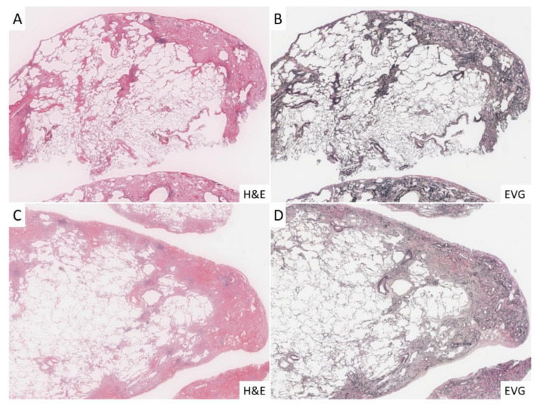
Figure 2.
Histopathological findings in the cases investigated for the current study. (A,B): In the PPFE group, fibroelastosis was significant under the pleura, and there was marked increase in elastic fibers with EVG staining. (C,D): In the IPF group, the development of dense fibrosis and fibroblastic foci was significant in subpleural and interlobular septa, and lung architecture destruction was highlighted with EVG staining.

