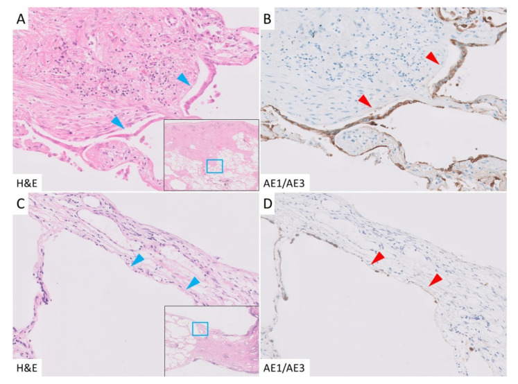
Figure 3.
Epithelial denudation in the PPFE group. (A): Alveolar epithelial denudation (AED) was detected in the border between the subpleural fibroelastosis and normal lung (blue arrowhead). (B): Cytokeratin AE1/AE3 immunohistochemical staining. The denudated epithelium and detached surface were highlighted in this staining (red arrowhead). (C,D): AED was also seen in the subpleural area without fibroelastosis (arrowhead).

