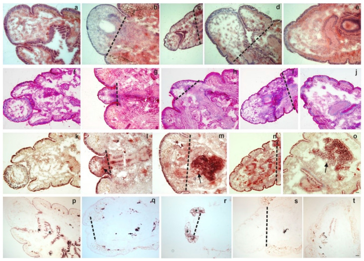
Figure 1.
Histochemistry of intact anterior segments (a,f,k,p) and regenerating blastema at various time points: 4 days (b,g,l,q); 1 week (c,h,m,r); 2 weeks (d,i,n,s) 4 weeks (e,j,o,t). Haematoxylin-eosin (H&E) (a–e), Periodic acid and Schiff reaction (PAS) (f–j), acid phosphatase (ACP) (k–o) and alkaline phosphatase (ALP) (p–t) stainings were performed. Representative images were presented from five independent experiments. Arrows point to ACP (m–o) positive cells. Asterisks mark ALP (q–r) expressing cells. The level of amputation is indicated by a dashed line. Scale bars: 200 μm.

