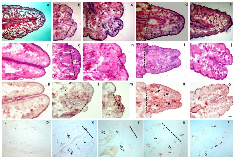
Figure 2.
Histochemistry of intact posterior segments (a,f,k,p) and regenerating blastema at various time points such as 4 days (b,g,l,q); 1 week (c,h,m,r); 2 weeks (d,i,n,s); 4 weeks (e,j,o,t). Standard histochemical staining methods were performed: (a–e) H&E, (f–j) PAS, (k–o) ACP and (p–t) ALP. In H&E images arrowheads point to coelomocytes (c–e) in the blastema. Representative images were selected from five independent experiments. Arrows point to ACP (l–n) expressing cells. Asterisks represent cells with ALP (q–t) expression. The level of amputation is denoted by a dashed line. Scale bars: 200 μm.

