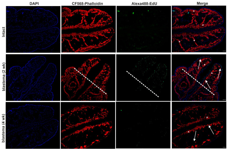
Figure 4.
Cell division and tissue re-organization in intact earthworms and in 2- and 4-week posterior blastema of regenerating earthworms. Representative images were chosen from five independent experiments. Arrows point to EdU-positive cells (green) and asterisks remark the actin filaments (red) in the blastema. The level of amputation is denoted by a dashed line. Scale bars: 200 μm.

