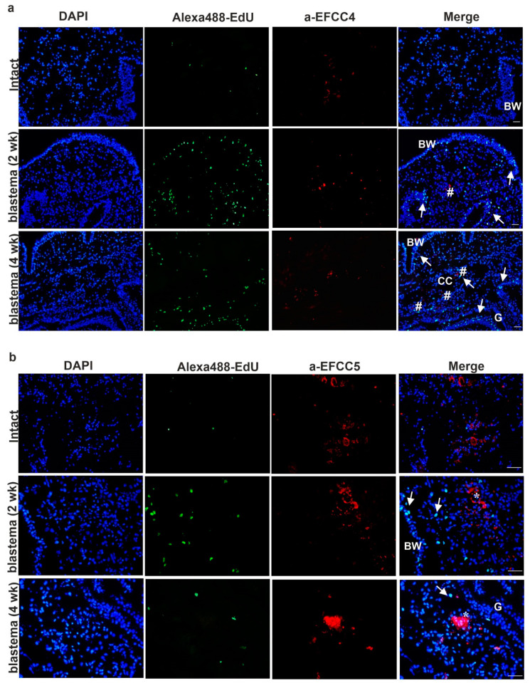
Figure 5.
Detection of cell division and localization of coelomocytes by specific mAbs in intact earthworms and 2- and 4-week anterior blastema: (a) granular amoebocytes (EFCC4-positive cells) and (b) eleocytes (EFCC5-positive cells). Arrows point to proliferating EdU-positive cells (green). (a) Number signs mark granular amoebocytes (red) and (b) asterisks denote eleocytes (red). Representative images were selected from five independent experiments. BW—body wall; CC—coelomic cavity; G—gut. Scale bars: 100 μm.

