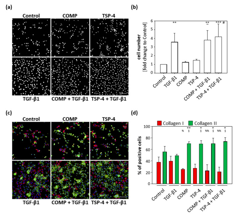
Figure 4.
Effect of COMP and TSP-4 on chondrocyte proliferation and dedifferentiation. Chondrocytes were stimulated with COMP (10 µg/mL) or TSP-4 (10 µg/mL) for seven days. (a) Nuclei were DAPI stained at day 10. (b) The fold changes of the counted nuclei are represented and each bar shows the mean + SD. The significant differences between the control and all other conditions were calculated. Differences between COMP, TGF-β1 and COMP + TGF-β1 as well as TSP-4, TGF-β1 and TSP-4 + TGF-β1 were calculated. Differences to the control are indicated by an asterisk *, to COMP by waves ~ and to TSP-4 by rhombus #, as well as significance indicated by p ≤ 0.05 #~; p ≤ 0.01 **; p ≤ 0.001 ***. (c) Immunofluorescence staining of collagen I (red), collagen II (green) and nuclei (blue) at day 10. (d) Percentages of chondrocytes expressing collagen I and II are represented and each bar shows the mean + SD. The significant difference between collagen I and collagen II positive cells was calculated and significance indicated as p ≤ 0.05 *; p ≤ 0.01 **. Differences of collagen I and collagen II positive cells to the control were calculated and significance indicated as p ≤ 0.05 % or §; p ≤ 0.01 %%, respectively. TGF-β1 (0.5 ng/mL) served as an inducer of proliferation and collagen synthesis and unstimulated cells as a control. (n = 3); scale bar = 100 µm.

