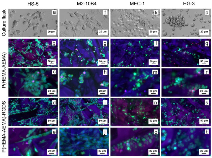
Figure 3.
Confocal micrographs of P(HEMA-AEMA)-RGDS scaffolds seeded with (a–e) HS-5, (f–j) M2-10B4, (k–o) MEC-1, (p–t) HG-3 cell culture for 24 h. (a,f,k,p) Transmitted light; (b–e, g–j, l–o, q–t) live and dead cells and nuclei stained by calcein acetoxymethyl ester (AM) (green), propidium iodide (red), and Hoechst 33342 (blue), respectively.

