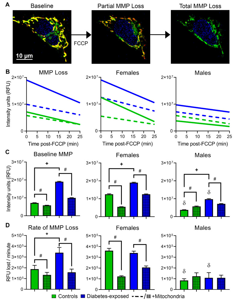
Figure 5.
Females and PGDM-exposed cells have stronger mitochondrial membrane potential (MMP), but they lose it faster following metabolic stress. (A) Representative images of CM stained with MitoTracker Green, Hoechst, and MitoTracker Red. Once treated with FCCP, mitochondria lose their MMP and trigger cell death through intrinsic apoptosis. (B) MMP loss was analyzed before and following FCCP-induced stress by linear regression analysis. (C) Baseline MMP and (D) rate of MMP loss over 25 min of FCCP-induced stress. N = 4–5 per sex per group. Data represent mean ± SEM. P ≤ 0.05: + diabetes, # mitochondrial, or δ sex-specific effect (only lower sex marked) by 1-way ANOVA.

