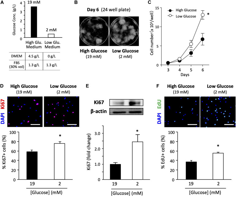
FIGURE 1.
Low-glucose medium increases the proliferation of primary satellite cells. (A) Glucose concentration in each growth medium used in this study. (B) Proliferation of primary satellite cells in high- and low-glucose media. Satellite cells from 20 myofibers were isolated from EDL and seeded in 24-well plates. Cell nuclei were visualized using DAPI and marked by the Hybrid Cell Count application (Keyence software). All the cells cultured in each well were automatically counted. (C) Cell growth curves. Values are presented as the mean ± SEM (n = 7). ∗p < 0.05. (D) Immunofluorescence analysis of proliferating cells cultured for 6 days. The population of Ki67-positive cells was quantified in high- and low-glucose media. Scale bars are 100 μm. Values are presented as the mean ± SEM (n = 3). ∗p < 0.05. (E) Western blot analysis of Ki67 protein expression in high- and low-glucose media after 6 days of cultivation. Ki67 expression was normalized to that of β-actin. Values are presented as mean ± SEM (n = 13). (F) Representative images of EdU+ satellite cells and the quantification of the number of EdU+ cells cultured for 6 days in high- and low-glucose media. Scale bars are 100 μm. Values are presented as the mean ± SEM (n = 4). ∗p < 0.05.

