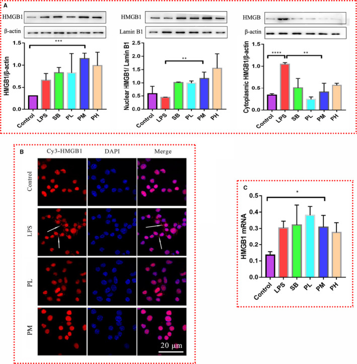
Figure 2.

Paeonol inhibits the translocation of HMGB1 from the nucleus to the cytoplasm. A, Total protein (left), nuclear protein (middle) and cytoplasmic protein (right) were extracted. The level of HMGB1 in each fraction was tested by using Western blotting. B, HMGB1 was stained with Cy3 (red), and nuclei were stained with DAPI (blue). HMGB1 was distributed in the cytoplasm (indicated by the white arrows). C, The relative mRNA expression level of HMGB1 was tested by using RT‐qPCR. The bar graphs are representative of three independent experiments. *P < .05, ** P < .01 and *** P < .001
