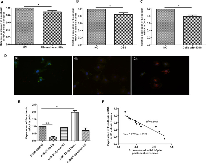
FIGURE 2.

E‐cadherin was down‐regulated in UC or DSS‐induced enteritis. E‐cadherin mRNA decreased in (A) intestinal mucosa of UC patients (n = 30), (B) intestinal mucosa of DSS‐induced enteritis mice (n = 15) and (C) DSS‐treated colonic epithelial cells. (D) Microscopic analysis of the internalization of the pre‐labelled exosomes in different time (0h, 4h, 12h). The exosomes displayed red colour. Cytoskeleton was stained with in green, and the cell nuclei were stained in blue, 400×. (E)There was a negative correlation between miR‐21a‐5p and E‐cadherin in colonic epithelial cells with different expression of miR‐21a‐5p. (F) Correlation analysis between intraperitoneal macrophage miR‐21a‐5p and E‐cadherin in intestinal mucosa from DSS‐induced enteritis mice (n = 15). The slope is −0.2733. R2 = 0.8464. * P < 0.05, ** P < 0.01. HC: healthy volunteers control group
