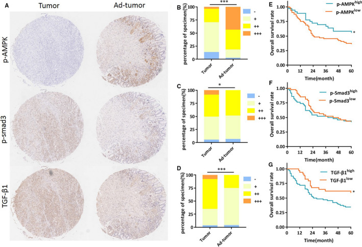
FIGURE 6.

TGF‐β1 level is decreased in gastric cancer and correlated with AMPK activation. A, Representative images of p‐AMPK (Th172), p‐Smad3 (Ser423/425) and TGF‐β1 in gastric carcinoma and adjacent tissues. B‐D, Statistical results of p‐AMPK (Th172), p‐Smad3 (Ser423/425) and TGF‐β1 were evaluated, respectively, according to Germany semi‐quantitative method and plotted. Evaluation of immunochemistry data showed that p‐AMPK was less than adjacent tumour tissues (P < .001), while p‐Smad3 and TGF‐β1 were higher in tumour tissues (P < .05 and P < .001 respectively). E‐G, Kaplan‐Meier survival curve for 5 year overall survival (OS) was assessed as opposed to on protein expression levels. OS for patients with higher p‐AMPK level was 55.6%, compared to 35.9% with lower p‐AMPK level (P < .05). OS for patients with higher TGF‐β1 level was 34.9%, compared to 51.4% of lower TGF‐β1 level (P < .05). No significance of OS was found with p‐Smad3 (P > .05)
