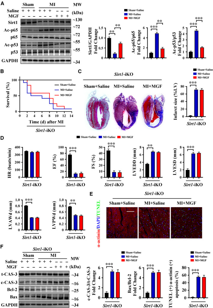
FIGURE 3.

Sirt1 is essential for the protective effects of mangiferin during MI. A, C57BL/6 mice were subjected to sham or LAD ligation followed by the treatment with or without mangiferin for 14 d, Western blotting assay and quantitative analysis of Sirt1, acetylated p65 (Ac‐p65), p65, acetylated p53 (Ac‐p53) and p53 in the heart tissue homogenates of different groups of mice. B‐F, Sirt1‐iKO mice were subjected to sham or LAD ligation followed by the treatment with or without mangiferin for 14 d. B, Kaplan‐Meier survival curves of different groups of mice (n = 15 mice in each group). C, Representative images of Masson's trichrome staining of the heart sections and macroscopic measurements of the infarct size. D, M‐mode echocardiography data for HR, EF (%), FS (%), LVEDD, LVESD, LVAWd and LVPWd. E, Representative confocal scans for α‐actinin, TUNEL and DAPI staining (red, green and blue, respectively) of the heart sections and quantitative analysis (lower panel) of TUNEL + and α‐actinin + cells, Scale bar = 100 μm. F, Western blotting assay and quantitative analysis of cleaved caspase 3 expression and Bax/Bcl‐2 ratio in the heart tissue homogenates of different groups of mice. Unless otherwise stated, data are mean ± SEM for n = 6 mice in each group. *P < .05; **P < .01; ***P < .001
