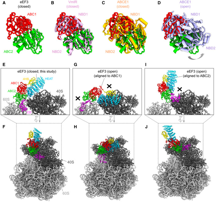
Figure EV3. The closed and open conformations of eEF3.

-
AThe ABC1 (red) and ABC2 (green) domain of eEF3 in the eEF3‐80S complex.
-
B–DConformation of the eEF3 NBDs with respect to other ABC proteins. Alignment (based on ABC1) of the eEF3‐ABCs with (B) the closed conformation of the NBDs of the B. subtilis ABCF ATPase VmIR (pink, PDB: 6HA8) (Crowe‐McAuliffe et al, 2018), (C) the closed conformation of the archaeal ABCE1‐30S post‐splitting complex (orange, PDB: 6TMF) (Nurenberg‐Goloub et al, 2020), and (D) the E. coli ABCE1 protein observed in the open conformation (violet, PDB ID: 3OZX) (Barthelme et al, 2011).
-
E, FThe eEF3 model in a closed conformation colored due to its different domain organization.
-
G–JIncompatibility of eEF3 to the 80S ribosome in a potential opened conformation. The eEF3 model was aligned to (G, H) ABC1 or (I, J) ABC2 of the ABCE1 protein in an opened conformation (PDB ID: 3OZX) (Barthelme et al, 2011).
