-
A
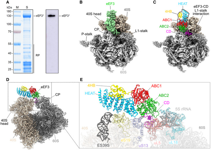
SDS gel and Western blot of the native pull‐out from S. cerevisiae TAP‐tagged eEF3. eEF3* labels the TEV cleavage product of eEF3‐TAP, which still carries the calmodulin binding peptide of the TAP‐tag and which is recognized by the antibody. RP, ribosomal proteins.
-
B
Cryo‐EM reconstruction of the native eEF3‐80S complex with segmented densities for the 60S (gray), the 40S (tan), and eEF3 (pastel green).
-
C
Fitted eEF3 molecular model colored according to its domain architecture, HEAT (blue), 4HB (yellow), ABC1 (red), ABC2 (green), and CD (magenta).
-
D, E
(D) Overview on the eEF3‐80S molecular model and (E) zoom illustrating eEF3 and its ribosomal interaction partners. ES39S (dark gray), eS19 (pale yellow), uS13 (pastel violet), uL5 (coral), uL18 (light blue), 5S rRNA (light gray).

