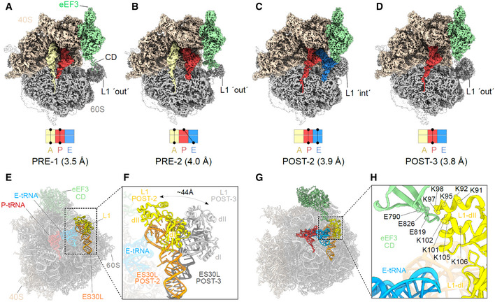
Figure 5. eEF3 bound to a non‐rotated 80S ribosome.

-
A–DCryo‐EM maps of eEF3 bound to non‐rotated ribosomal species with isolated densities for the 60S, 40S and eEF3 (colored as in Fig 1), as well as the A‐ (pale yellow), P‐ (red), and E‐tRNA (blue). The eEF3 ligand is binding the ribosome in the pre‐translocational (PRE) state termed as (A) PRE‐1 (A/A‐, P/P‐tRNA) and (B) PRE‐2 (A/A‐, P/E‐tRNA) as well as to the post‐translocational (POST) version named (C) POST‐2 (P/P‐, E/E‐tRNA) and (D) POST‐3 (P/P‐tRNA).
-
E, F(E) Overview of the POST‐2 eEF3‐80S molecular model highlighting the L1 protein (yellow) and ES30L (orange) and its (F) zoom depicting the magnitude of the L1‐stalk movement from the POST‐2 state (L1‐stalk′ int′) to the POST‐3 state (L1‐stalk′out′). L1‐POST3 (light gray), ES30L‐POST3 (dark gray).
-
GDifferent view of the model shown in (E) highlighting eEF3, L1, ES30L as well as the P/P‐ and E/E‐tRNA.
-
HZoom of (G) showing the contact of the eEF3‐CD and the L1 protein as well as the distance of both to the E‐tRNA. The residues are displayed as circles and labeled.
