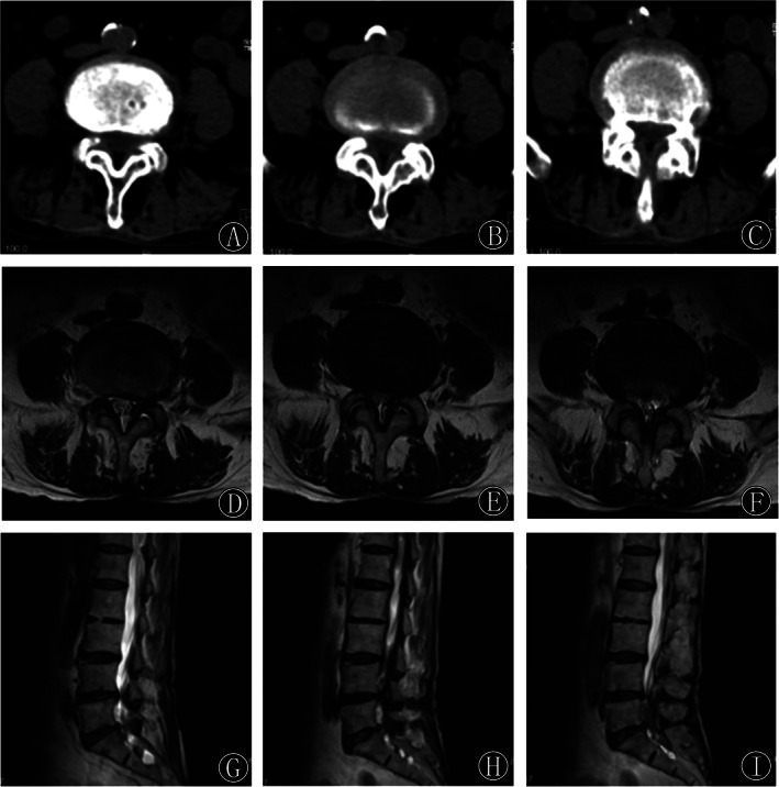
Fig. 1.

(A–C) Preoperative consecutive axial CT images showed facet joint hyperplasia and cohesion at the L4‐5 segment, resulting in spinal canal stenosis and severe compression of the dural sac and spinal roots. (D–I) Preoperative axial and sagittal T2‐weighted MRI images showed spinal stenosis at the L4‐5 segment with significant compression of the dural sac.
