
Fig. 4.
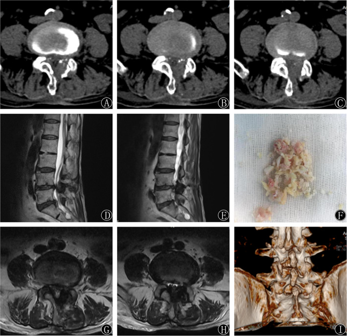
(A‐H) Sagittal MRI, axial MRI, and CT showed satisfactory decompression effects 1 day postoperatively. The medial edge of the lamina, hypertrophy of the ligamentum flavum, and articular process were removed, and the spinal cord was decompressed. (I) A round‐shaped laminotomy at the L4‐5 segment was shown on the 3D reconstruction of the CT scan.