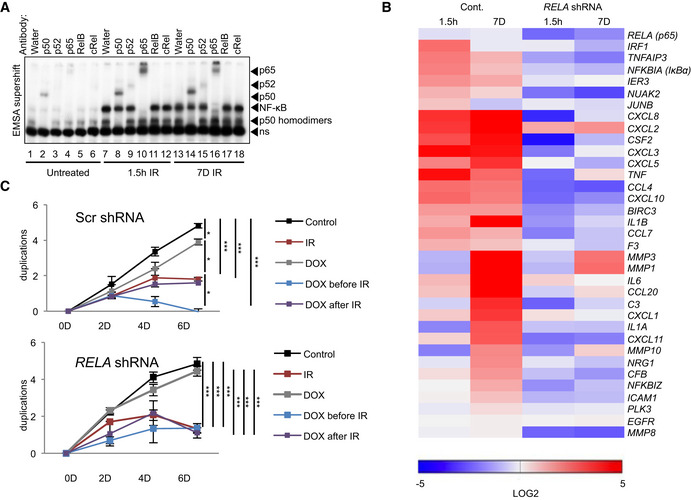
Figure 3. RelA regulates SASP but not cell cycle arrest.

- Nuclear fractions from either untreated or irradiated (20 Gy) U2‐OS cells, 1.5 h or 7 days prior to harvest, were analyzed by EMSA with supershift analysis. For supershifting, lysates were incubated with antibodies or water, as indicated. Arrows point to shift location of a given subunit, due to antibody binding. ns, non‐specific band. All antibodies were previously tested for supershift compatibility. Representative gel shown from n = 3 replicates.
- Expression of SASP targets of NF‐κB obtained from RNA‐seq analysis (Dataset EV1B), was quantitated using RT–qPCR from the shRNA (RELA) stably expressing U2‐OS cells. For knockdown efficiency, see Appendix Fig S4A. Cells were treated with doxycycline (Dox) to induce knockdown of p65. Heatmap represents targets normalized to the untreated Scrambled control. Expression is shown as log2 change with P values < 0.05. Statistical analysis performed using ANOVA with Bonferroni correction for multiple testing. First‐phase and second‐phase samples were irradiated (20 Gy) 1.5 h and 7 Days prior to harvest, respectively. Analysis based on n = 3 biological replicates.
- Cell duplication was measured at time points indicated in U2‐OS cells bearing either scrambled control or Dox‐inducible shRNA against RELA (n = 3 biological replicates). Treatment with Dox was initiated at 2 days prior to IR (20 Gy) or after IR. Statistical significance in total duplication number between groups at day 6 was determined by ANOVA with Tukey multiple comparisons test. SD shown. *P < 0.05, ***P < 0.001.
Source data are available online for this figure.
