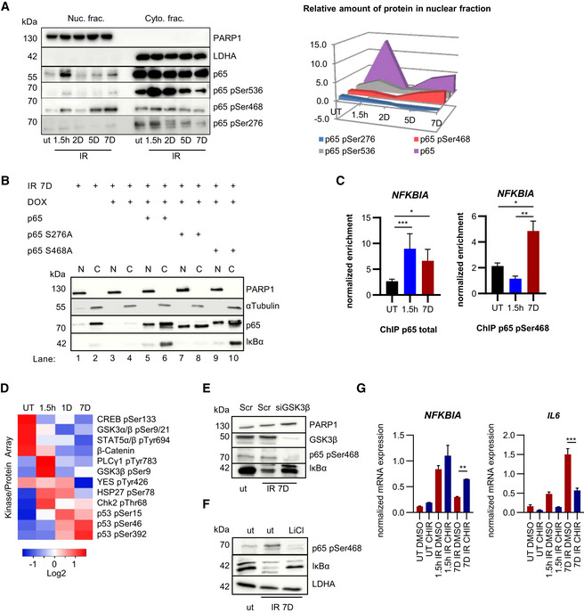
Figure 4. p65 Ser 468 phosphorylation in senescence inhibits NFKBIA expression.

- Nuclear (Nuc.) and cytoplasmic (Cyto.) fractions of U2‐OS cells were analyzed at times indicated after IR exposure by SDS–PAGE Western blotting. PARP1 and LDHA are fractionation controls and phospho‐specific p65 signals are indicated. Representative gels from n = 3 biological replicates are shown. Right panel: quantitation of nuclear p65 and phosphorylated p65 species (n = 3). Fold changes compared to untreated samples (ut) and time points post‐IR are indicated on Y and X axes, respectively.
- U2‐OS cells treated with Dox to deplete endogenous p65 were irradiated (20 Gy) and transfected with plasmids encoding p65, p65‐S276A or p65‐S468A, as indicated. Nuclear (N) and cytoplasmic (C) lysates were analyzed by SDS–PAGE at day 7 post‐IR. Representative gel from n = 3 biological replicates is shown. PARP1 and α‐tubulin serve as fractionation and loading controls.
- ChIP performed with U2‐OS cells irradiated (20 Gy) 1.5 h or 7 days prior to assay, or left untreated (UT). Normalized relative enrichment of NFKBIA is shown (relative to input and two non‐recruiting reference regions). Left panel: p65 antibody. n = 5 biological replicates with three technical repeats per biological replicate. Right: Antibody against p65 pSer468. n = 2 biological replicates with three technical repeats per biological replicate. Statistical significance was determined by ANOVA with Tukey multiple comparisons test. SD shown. *P < 0.05, **P < 0.01, ***P < 0.001.
- Kinase proteome array was performed with nuclear fractions of U2‐OS cells that were either left untreated (UT) or irradiated (20 Gy) at time points indicated prior to harvest. Kinase binding to membrane was quantitated (n = 2) and only significant results (P < 0.05) are shown. Significance was determined by ANOVA with Tukey multiple comparisons test.
- U2‐OS cells left untreated (ut) or irradiated (20 Gy) 7 days before harvesting, as indicated. Two days prior to harvest cells were transfected with siRNA against GSK3β or scrambled control (Scr). Whole cell lysates were analyzed by SDS–PAGE and Western blotting. A representative example from n = 3 experiments is shown.
- U2‐OS cells were left untreated or exposed overnight to 10 mM LiCl, with or without prior irradiation, as indicated, and analyzed as in (E). Representative data from n = 3 experiments are shown.
- RT–qPCR of U2‐OS cells irradiated (20 Gy) either 1.5 h or 7 days prior to harvest. CHIR denotes treatment with GSK3 inhibitor CHIR‐99021 (10 nM overnight). DMSO overnight treatment was used as control. Quantitation was performed and statistical significance obtained from n = 3 replicates, using ANOVA with Tukey multiple comparisons test. SD shown. *P < 0.05, **P < 0.01, ***P < 0.001.
Source data are available online for this figure.
