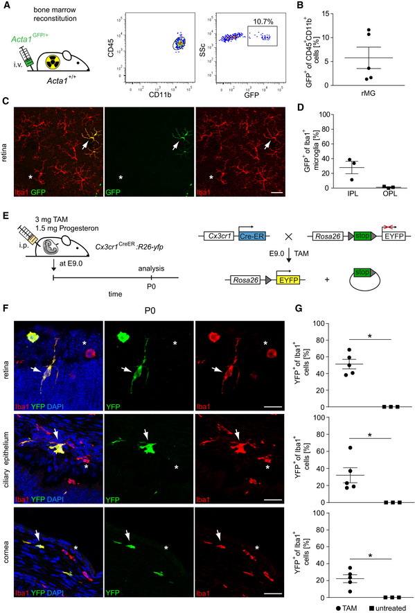
Left: Creation of Acta1
GFP/+
:Acta1
+/+ bone marrow chimeras. Right: Donor‐derived GFP+CD45loCD11b+ were detectable in the recipient retina by flow cytometry 20 weeks after bone marrow reconstitution. FACS Plots are representative for five animals from one experiment.
Quantification of GFP+ cells among CD45+CD11b+ retinal microglia by flow cytometry. Data are presented as mean ± s.e.m. One symbol represents one mouse.
Typical retinal flat mount from Acta1
GFP/+
:Acta1
+/+ bone marrow chimeras 20 weeks after reconstitution. Donor‐derived GFP+Iba1+ cells (arrow) and GFP−Iba1+ resident microglia (asterisks) are shown. Pictures are representative for three animals from one experiment. Scale bars represent 50 µm.
Microscopy‐based quantification of GFP+Iba1+ retinal microglia in the inner (IPL) and outer plexiform layer (OPL). One symbol represents one mouse. Data are presented as mean ± s.e.m.
Scheme of a fate mapping experiment using Cx3cr1CreERT2:Rosa26‐YFP female mice. Tamoxifen (TAM) and progesterone injection were performed at embryonic day 9.0 (E9.0). Mice were subsequently evaluated at postnatal day 0 (P0). Administration of TAM leads to intra‐embryonic excision of a stop sequence flanked by loxP sites (gray triangles) in Cx3cr1 expressing cells which causes stable and steady YFP expression under the control of the Rosa26 promotor.
Direct fluorescence microscopic visualization for YFP (green), the macrophage marker Iba1 (red) and DAPI for the nuclei (blue) at P0. YFP+Iba1+ double‐positive cells are marked by arrows. YFP−Iba1+ single‐positive cells are labeled by asterisks. Representative images out of five examined animals are shown. Scale bars represent 25 µm.
Quantitative analysis of regional YFP expression in Iba1+ macrophages in TAM‐induced and untreated Cx3cr1CreERT2:Rosa26‐YFP mice. Bars represent means ± s.e.m. Quantification was done from three (untreated) or five (TAM) mice obtained from one (untreated) or two (TAM) independent experiments. Level of significance determined by Mann–Whitney test between TAM and untreated revealed *P < 0.05 and Kruskal–Wallis test between retina, ciliary body, and cornea revealed *P = 0.0204.