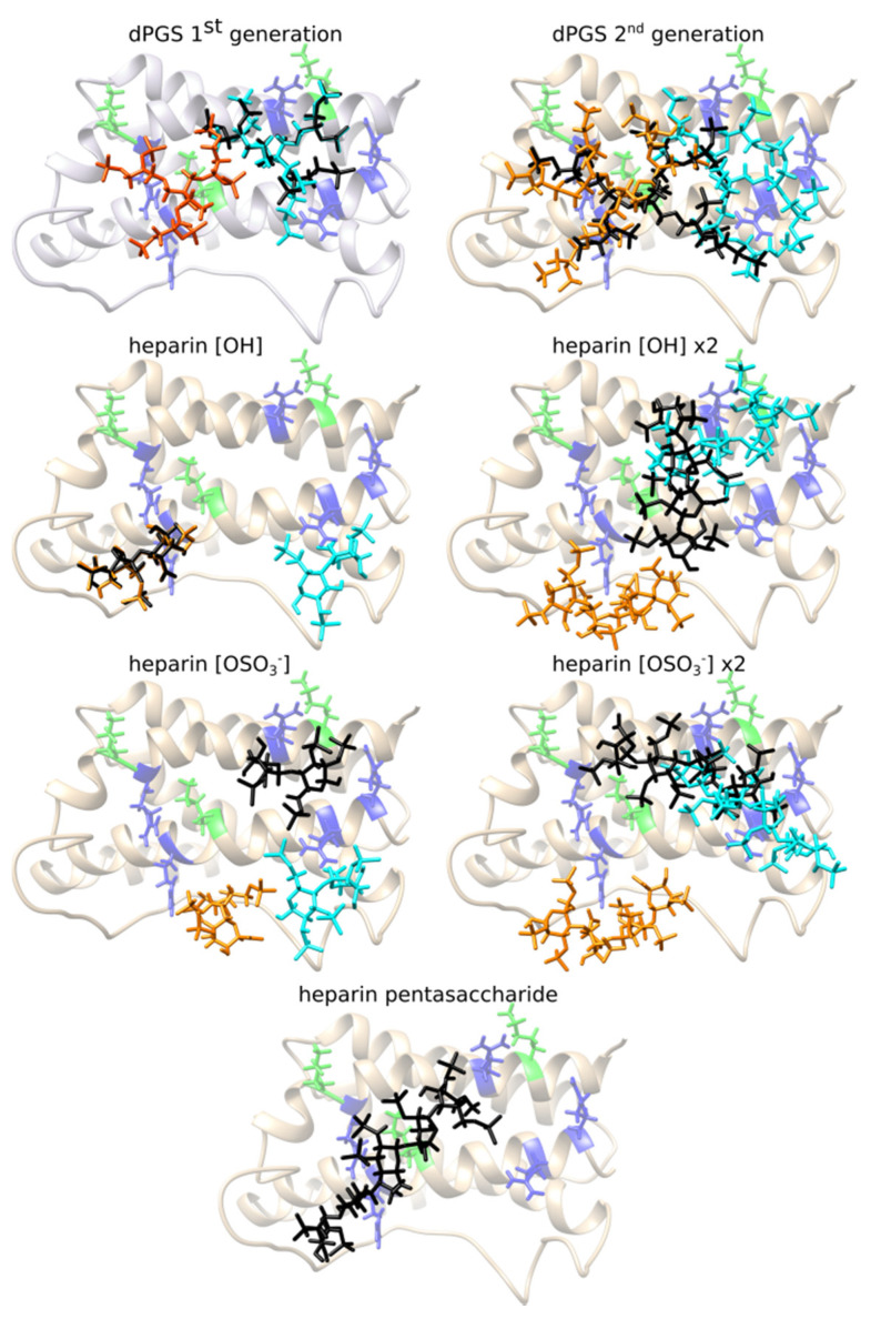
Figure 2.
The most favorable binding positions for different ligands and binding sites of IL-6. Lysine form the docking sites is colored green, arginine is colored blue. Orange structure represents the corresponding pose obtained in the RKRK docking box; cyan pose obtained in the KRRR docking box, and black is the pose obtained in the docking box (iii) RKRK&KRRR which surrounds both binding sites.

