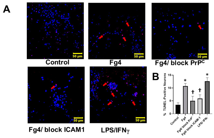
Figure 5.
Fibrinogen (Fg)-induced astrocyte activation causes apoptosis of co-cultured neurons (A) Representative images of neuronal apoptosis assessed by Terminal deoxynucleotidyl transferase-dUTP nick end labeling (TUNEL) assay. TUNEL assay is a method for detecting DNA fragmentation by labeling the 3′- hydroxyl termini in the double-strand DNA breaks generated during apoptosis. Neurons were co-cultured with astrocytes treated with Fg for 24 h. Astrocytes were treated with medium alone (control), 4 mg/mL of Fg (Fg4), 4 mg/mL of Fg in the presence of a function-blocking peptide against cellular prion protein (Fg4/block PrPC) and 4 mg of Fg in the presence of function-blocking antibody against intercellular adhesion molecule-1 (Fg4/block ICAM-1). For positive control, lipopolysaccharide (LPS) was used at 1µg/mL with co-stimulation with 20 ng of a murine interferon gamma (IFNγ). Immunostaining of apoptotic (red) neurons with 4′,6-diamidino-2-phenylindole (DAPI, blue) nuclear stain is shown. Arrows indicate some of the apoptotic cells (red). (B) Summary of image analyses for the detection of apoptotic neurons presented as a percent of a total number of cells. p < 0.05 in all; *—vs. Control, †—vs. Fg4; n = 4.

