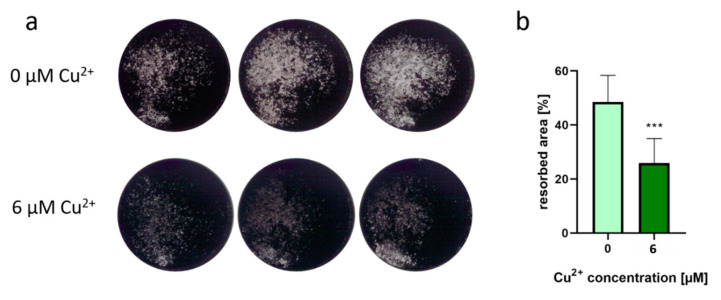
Figure 6.
(a) Representative images of osteoblast-derived ECM after osteoclastic resorption in the presence of 6 µM Cu2+ compared to a copper-free control. SaOS-2 osteoblasts were cultivated for 4 weeks until a closed layer of mineralized extracellular matrix was formed in the dishes. After removal of the osteoblasts, PBMC (two donors, each n = 4 per group) were seeded and cultivated for 14 days under stimulation with M-CSF and RANKL with and without addition of Cu2+. After fixing with 4% formaldehyde, von Kossa staining was performed to stain the remaining mineralized matrix after osteoclastic resorption. Images were recorded with a Leica stereomicroscope and represent the whole area of a 48- well dish (12 mm diameter). (b) Resorbed area of all samples was calculated applying the open source software Fiji using the threshold function. Eight samples were imaged for each condition and shown as mean +/− standard deviation. Significant differences were calculated by two-tailed unpaired t-test with p < 0.001.

