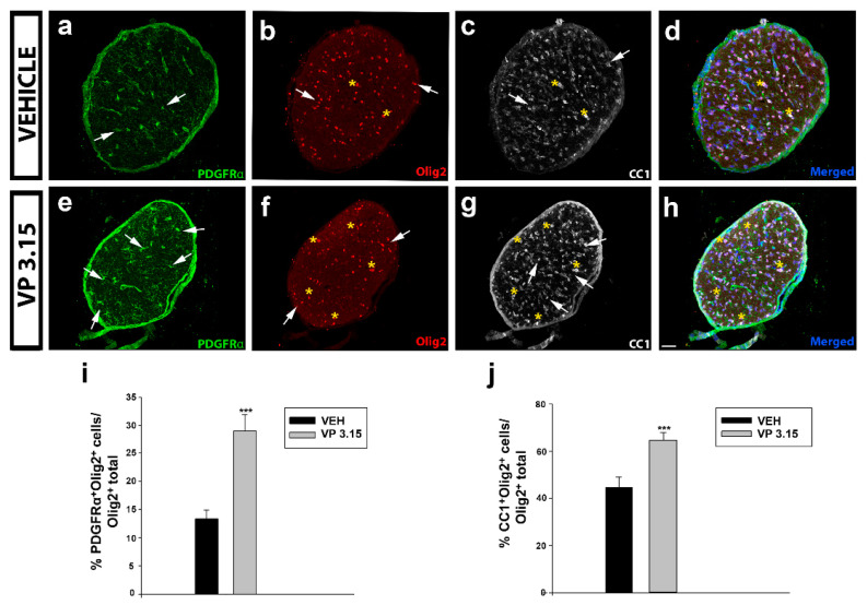
Figure 6.
VP3.15 promotes the presence of both precursor and mature oligodendrocytes in optic nerve; (a–h): Detailed views of the optic nerve of vehicle (a–d) and VP3.15-treated mice (e–h), labelled for PDGFRα (green), Olig2 (red), CC1 (grey), and merged includes nuclei (Hoechst; blue). Arrows point to PDGFRα+Olig2+ cells and asterisks to CC1+Olig2+ cells. Scale bar represents 50 µm in a–h; (i,j): Graphs showing the significant increase in the percentage of PDGFRα+Olig2+ cells (i) and CC1+ Olig2+ cells (j) after the treatment with VP3.15 compared to the vehicle. Results of Student’s t-test are represented as: *** p < 0.001. EAE-VEH: n = 7; EAE-VP3.15: n = 10.

