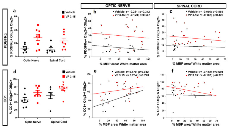
Figure 8.
The maturation state of oligodendrocytes is related to the myelin preservation in the optic nerve; (a–d): Graphs showing the different levels of precursor (PDGFRα+; a) and mature cells (CC1+ cells; d) between structures; (b,c): Graphs showing the correlation between the normalized MBP area and the percentage of PDGFRα+Olig2+ cells in both treatments and in the optic nerve (b) and spinal cord (c); (e,f): Graphs showing the results of Pearson´s correlation between the normalized MBP area and the CC1+ Olig2+ cells in both treatments and in the optic nerve (e) and spinal cord (f). EAE-VEH: n = 7; EAE-VP3.15: n = 10.

