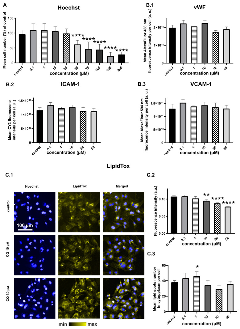
Figure 1.
HMEC-1 response to 24-h incubation with CQ: (A) Number of cells calculated based on Hoechst staining and presented as a % of the control for the nucleus number; (B.1) vWF; (B.2) ICAM-1; (B.3) VCAM-1 expression (ANOVA; * p-value < 0.05, ** p-value < 0.01, **** p-value < 0.0001); representative fluorescence images of cells stained with Hoechst and LipidTox (C.1) fluorescence intensity of lipid spots (C.2) and their mean number in the cytoplasm (C.3). CQ affected cell viability at 50 µM, had no effect on vWF, VCAM-1, ICAM-1 overexpression, and increased the neutral lipid content (ANOVA, p-value < 0.05). Average fluorescence value from six wells was quantified from >9000 cells per well. Results were obtained from three independent experiments.

