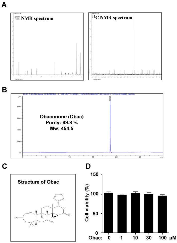
Figure 1.
Effects of Obac on proliferation in MC3T3-E1. (A) 1H and 13C NMR spectra of Obac from the dried root bark of Dictamnus dasycarpus Turcz. (B) HPLC chromatogram of Obac. (C) Chemical structure of Obac. (D) MC3T3-E1 was incubated with Obac at concentrations of 1, 10, 30, and 100 μM for 24 h, and cell viability was measured by the MTT assay. Data are representative of three independent experiments, and values are expressed in the mean ± S.E.M

