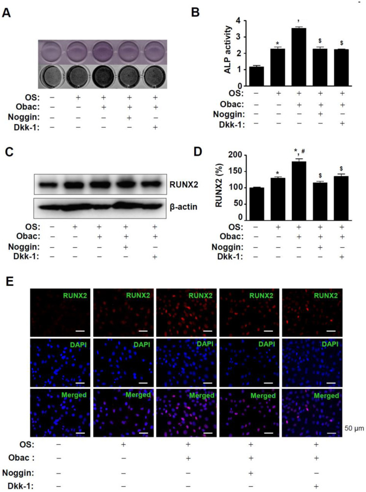
Figure 6.
Effects of Obac-induced osteoblast differentiation and RUNX2 expression through the inhibition of BMP2 and β-catenin signaling. (A,B) MC3T3-E1 was incubated in OS with 10 µM Obac in the absence or presence of noggin (10 µg/mL) and Dkk-1 (0.5 µg/mL) for 7 days. ALP staining was detected using a scanner (upper) and a colorimetric detector (bottom) (A), and ALP activity was assessed using ALP activity colorimetric assay (C). (C,D) After 48 h, the expression of RUNX2 was assessed using Western blot analysis (C). β-actin was detected on the same sample to normalize the values, and the data were expressed as a bar graph (D). (E) After 48 h, the nuclear localization of RUNX2 were assessed using Immunofluorescence. The first panels show RUNX2 expression (red), the second panels show DAPI (a nuclear marker, blue), and the bottom panels show the merged images of the first and second panels. Scale bar: 50 μm. Data are representative of three independent experiments, and values are expressed in the mean ± S.E.M. * p < 0.05 indicates statistically significant differences, compared with the control. #: statistically significant differences compared with OS (# p < 0.05). $: statistically significant differences compared with the OS + Obac ($ p < 0.05).

