
Figure 4. Inhibition of HSP90, but not HSP70, regulates the packaging of GOF p53 into small EVs.
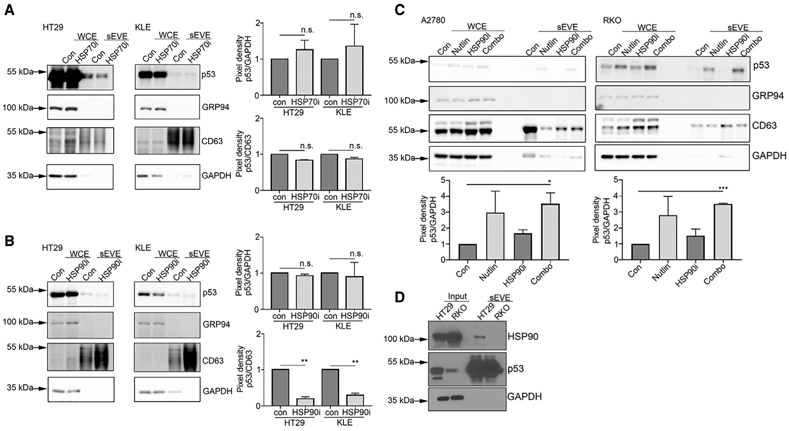
(A) WCE and sEVE were used to determine the level of p53 protein after treatment with the HSP70 inhibitor (HSP70i) VER-155008. Data are represented as mean ± SD. n.s., not significant.
(B) Expression of GOF p53 protein in small EVs in HT29 and KLE cell-derived sEVE after treatment with the HSP90 inhibitor (HSP90i) 17-AAG. Data are represented as mean ± SD. **p < 0.01; n.s., not significant.
(C) WT p53 protein expression in A2780 and RKO cells after treatment with nutlin-3A, an HSP90i, or a combination of the two. Pixel densities for all p53 bands in comparison with GAPDH and CD63 bands normalized to 1.0 are shown. Data are represented as mean ± SD. *p < 0.05; ***p < 0.001.
(D) Results of a co-immunoprecipitation assay performed using HT29 and RKO cells and their relatively small EVs.