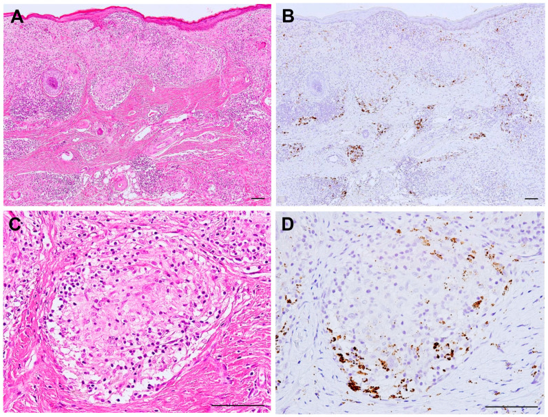
Figure 4.
Active P. acnes infection in granulomatous inflammation of cutaneous sarcoidosis. Hematoxylin–eosin stain and immunohistochemistry with P. acnes-specific PAB antibody are shown pairwise. Many granulomas are found in the dermis with prominent lymphocytic infiltration (A). Many PAB-reactive P. acnes (in brown color) are found corresponding to the areas of granulomatous inflammation (B). In a non-caseating epithelioid cell granuloma (C), P. acnes is abundant in the peripheral area with more lymphocytic infiltration (D). All photos are original. Scale bar: 100 μm.

