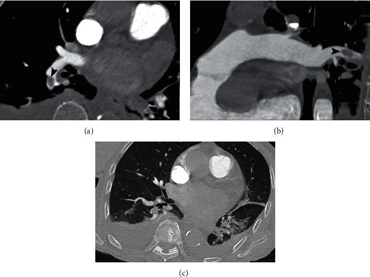
Figure 2.

CTPA of a 94-year-old woman with SARS-CoV-2 infection. (a) Axial image with mediastinal window. (b) Curved MRP image with mediastinal window. PAFD of the lobar artery for the right lower lobe (arrowhead). (c) Lung window shows linear opacities and consolidation in the left lower lobe and bilateral pleural effusion.
