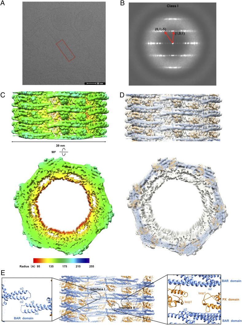
Fig. 2.
Cryo-EM and three-dimensional reconstructions of SNX1 coated tubules. (A) Raw cryo-EM micrographs of tubules coated with SNX1. A regular tube is boxed in red (scale bar, 50 nm). (B) Helical diffraction patterns of tubules exemplifying class I. (C) Cryo-EM map of a class I tubule, with the side view on the top and cross-section view on the bottom, with a lower threshold in cross-section view to show the inner membrane more clearly. The map is colored according to the cylinder radius from red to blue. (D) The structural models of SNX1 in cartoon representation are fitted into the map. The PX domain and BAR domain are colored in gold and blue, respectively. (E) Structural model of the SNX1 helical assembly for the class I tubules. Interface I and II are indicated by black circles, with zoom-in views also shown.

