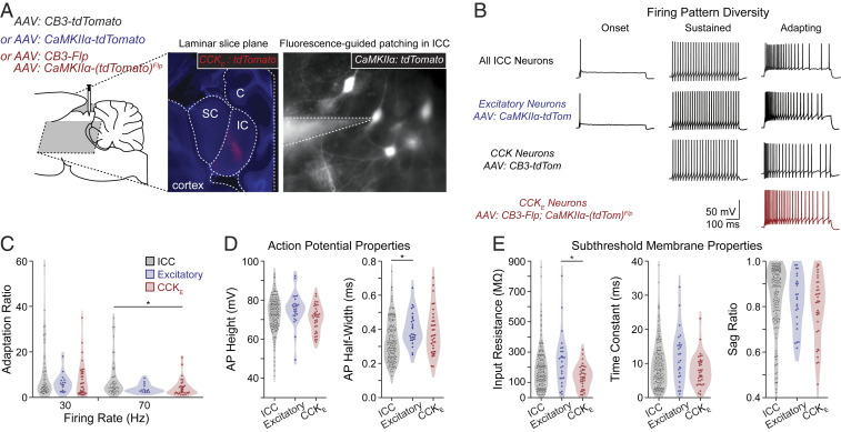
Fig. 5.
CCKE neurons have an adapting firing pattern in vitro. (A) AAV injections were made in the gerbil ICC. Middle shows a representative slice containing viral targeted CCKE neurons (red) and counterstained for DAPI after fixation. Right shows a recording of a viral targeted excitatory neuron. SC: superior colliculus; IC: inferior colliculus; C: cerebellum. (B) Whole-cell current-clamp recordings were made of unlabeled or virus-targeted fluorescent IC neurons. Unlabeled and virus-targeted excitatory neurons displayed the full range of firing pattern diversity, including adapting, sustained, and onset firing patterns (unlabeled: n = 68, 79, and 5/152 neurons, respectively, n = 83 gerbils; labeled excitatory: n = 16, 6, and 4/26 neurons, respectively, n = 18 gerbils). Labeled CCK neurons had only adapting and sustained firing patterns (34 and 5/39 neurons, respectively, n = 18 gerbils). Labeled CCKE neurons all had adapting firing patterns (n = 35/35 neurons, n = 21 gerbils). Sustained and adapting examples had firing rates of ∼50 Hz. (C) The adapting firing pattern of CCKE neurons was not different from that of other ICC neurons displaying adapting firing. Adaptation ratios were measured for unlabeled and fluorescence-targeted (all excitatory or CCKE) neurons for firing rates from bins of 20 to 40 and 60 to 80 Hz. The following are reported as mean ± SEM. Adaptation ratio at 30 Hz: all ICC = 9.9 ± 1.6; all excitatory = 6.3 ± 1.0; CCKE = 7.7 ± 1.3. Adaptation ratio at 70 Hz: ICC = 8.1 ± 1.2; all excitatory = 3.9 ± 0.5; CCKE = 4.4 ± 0.7. Sample sizes were ICC: n = 53 cells, n = 41 gerbils; all excitatory: n = 16 neurons, n = 13 gerbils; CCKE: n = 35 neurons, n = 21 gerbils. ANOVA and subsequent Tukey test resulted in one comparison with P < 0.05 (CCKE and ICC at 70 Hz, P = 0.043). (D and E) Active and subthreshold membrane properties of CCKE neurons were not different from those of other ICC neurons. Measurements were made from unlabeled and fluorescent excitatory and CCKE neurons. The following are reported as mean ± SEM. AP height: ICC = 72.95 ± 0.73 mV; all excitatory = 75.45 ± 1.64 mV; CCKE = 70.78 ± 1.14 mV. AP half-width: ICC = 0.33 ± 0.01 ms; all excitatory = 0.40 ± 0.02 ms; CCKE = 0.38 ± 0.02 ms. Input resistance: ICC = 195.8 ± 11.7 MΩ, all excitatory = 246.9 ± 34.0 MΩ, CCKE = 138.0 ± 12.8 MΩ. Time constant: ICC = 9.5 ± 0.5 ms; all excitatory = 12.1 ± 1.5 ms; CCKE = 8.0 ± 0.8 ms. Sag ratio: ICC = 0.84 ± 0.01; all excitatory = 0.83 ± 0.02; CCKE = 0.80 ± 0.02. ANOVA and subsequent Tukey test resulted in two comparisons with P < 0.05 (AP half-width for ICC and excitatory, P = 0.014; input resistance for excitatory and CCKE, P = 0.008).

