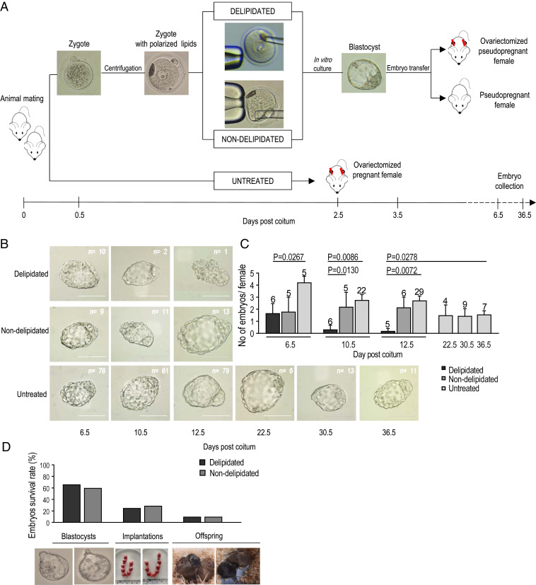
Fig. 1.
Delipidated embryos are unable to survive during the progression of ED. (A) Workflow of the generation of delipidated and nondelipidated embryos (control manipulation—removal of cytoplasm instead of lipids), their transfer to pseudopregnant ovariectomized females (number of transferred embryos per female = 16), and subsequent collection. Untreated embryos were developed entirely in vivo in ovariectomized females. (B) Representative images of blastocysts collected at progressive days of ED show temporal survival of delipidated and control (nondelipidated, untreated) embryos during ED. The proportions of collected delipidated and nondelipidated embryos were similar: 10 of 96 (10.42%) vs. 9 of 80 (11.25%) at 6.5 dpc. Then, they reduced drastically for delipidated embryos: 2 of 96 (2.08%) vs. 11 of 80 (13.75%) at 10.5 dpc and 1 of 80 (1.25%) vs. 13 of 96 (13.54%) at 12.5 dpc. (Scale bars, 100 μm.) (C) Histogram shows that only 3 (1.7%) delipidated embryos of 176 transferred were still present in the uterus of ovariectomized females at 10.5 and 12.5 dpc. At the same time, untreated embryos were collected from the uteri of ovariectomized mice without any significant decline in their survival until 36.5 dpc, which implies a crucial role of the lipid fraction in ED extension (demonstrated in the last part of this study) (Fig. 4). Numbers above the columns indicate the numbers of females carrying diapausing embryos. Values represent mean ± SEM; Mann–Whitney test. (D) Graph and representative images (Lower) demonstrate similar survival of delipidated and nondelipidated embryos at blastocyst stage (123 of 206 vs. 85 of 128) and following implantation (21 of 72 vs. 14 of 57) and delivery (3 of 30 vs. 2 of 20; χ2 test, no significant differences).

