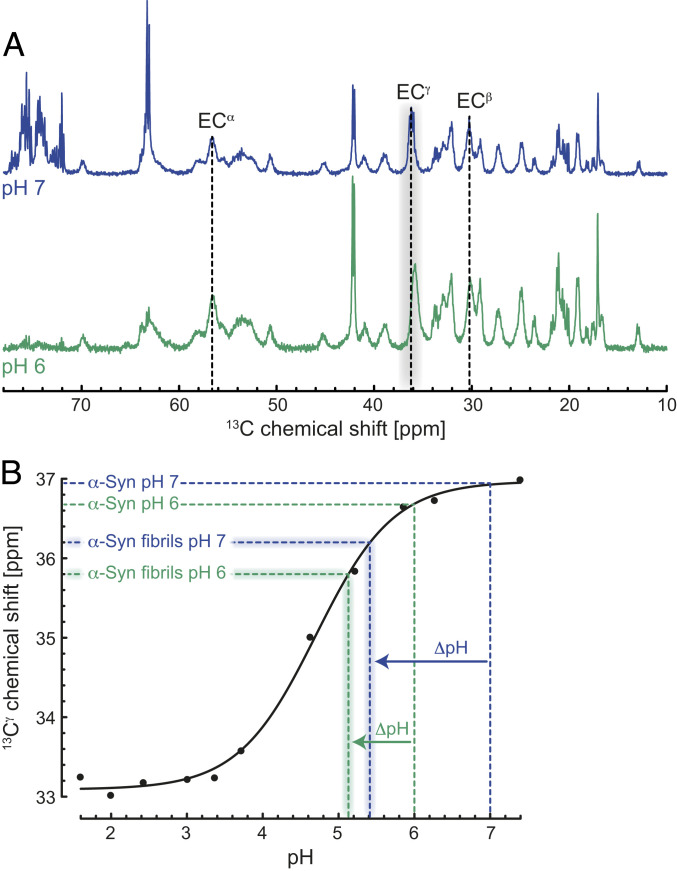
Fig. 4.
Lowered local pH on the surface of the α-Syn fibrils triggers monomer–fibril interactions. (A) One-dimensional (1D) 13C MAS INEPT NMR spectra of the mobile regions of α-Syn fibrils at pH 7 (blue) and 6 (green) in the absence of salt. 13C chemical shifts of Glu residues are shown in one-letter amino acid code. (B) Comparison of the pH titration data reported for Glu114 13Cγ of soluble α-Syn (black, reproduced from ref. 39) and the Glu 13Cγ chemical shifts of the α-Syn fibrils obtained from the 1D 13C INEPT spectrum. The local ΔpH differences between soluble and α-Syn fibrils at pH 7 (blue) and 6 (green) are indicated.

