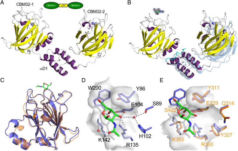
Fig. 3.
Structures of the family 32 CBMs. (A) Modular schematic of the crystallized and modeled CBM32-1/2 protein according to Fig. 1 shown with a cartoon representation of the CBM32-1/2 crystal structure. The N and C termini are indicated and bound calcium ions are shown as blue spheres. (B) Cartoon representation of the CBM32-1/2 structure in complex with GalNAc (shown as green sticks). The electron density for the GalNAc residue is shown as a σa-weighted Fo-Fc omit map contoured at 3σ. A peptide derived from the N terminus of the protein that was bound at an interface of CBM32-1 and αD1 is shown as a cyan ribbon. This structure was overlayed via the CBM32-1 modules with the CBM32-1/2 structure determined in the absence of GalNAc; the unbound structure is shown in transparent blue. (C) Overlap of the CBM32-1 module (blue with GalNAc as green sticks) with the CBM32-2 (orange) from the complexed structure. (D) Close-up of the CBM32-1 binding site showing specific interactions and the solvent accessible surface as transparent gray. (E) An overlap of the CBM32-1 binding site (blue) with the analogous region of CBM32-2 (orange with orange labels). The surface of CBM32-2 is shown as transparent gray.

