Fig. 4.
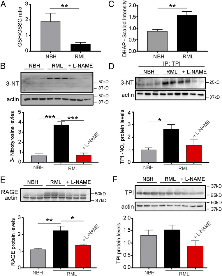
NOS inhibition reduces protein 3-nitrotyrosination and glycation signaling. (A) The ratio of reduced/oxidized glutathione is diminished in hippocampal tissue from RML mice at 10 w.p.i.. (B) Levels of 3-NT proteins are enhanced in RML and reduced following L-NAME treatment of RML mice. (C) DHAP levels are increased in the hippocampus of RML mice. (D) The 3-NT formation of triose-phosphate isomerase (TPI) is increased in RML mice and reduced to control NBH levels following L-NAME treatment of RML mice. (E) RAGE levels (upper band; lower band represents actin) are enhanced in RML hippocampi. (F) Total TPI levels are not affected by prion disease or treatment. Data are presented as mean ± SEM, n = 6 NBH, n = 9 RML mice (A and C) and n = 3 mice each in B and D–F. Unpaired Student’s t test, **P < 0.01 (A and C), one-way ANOVA, *P < 0.05, **P < 0.01, ***P < 0.001 (B and D–F)

