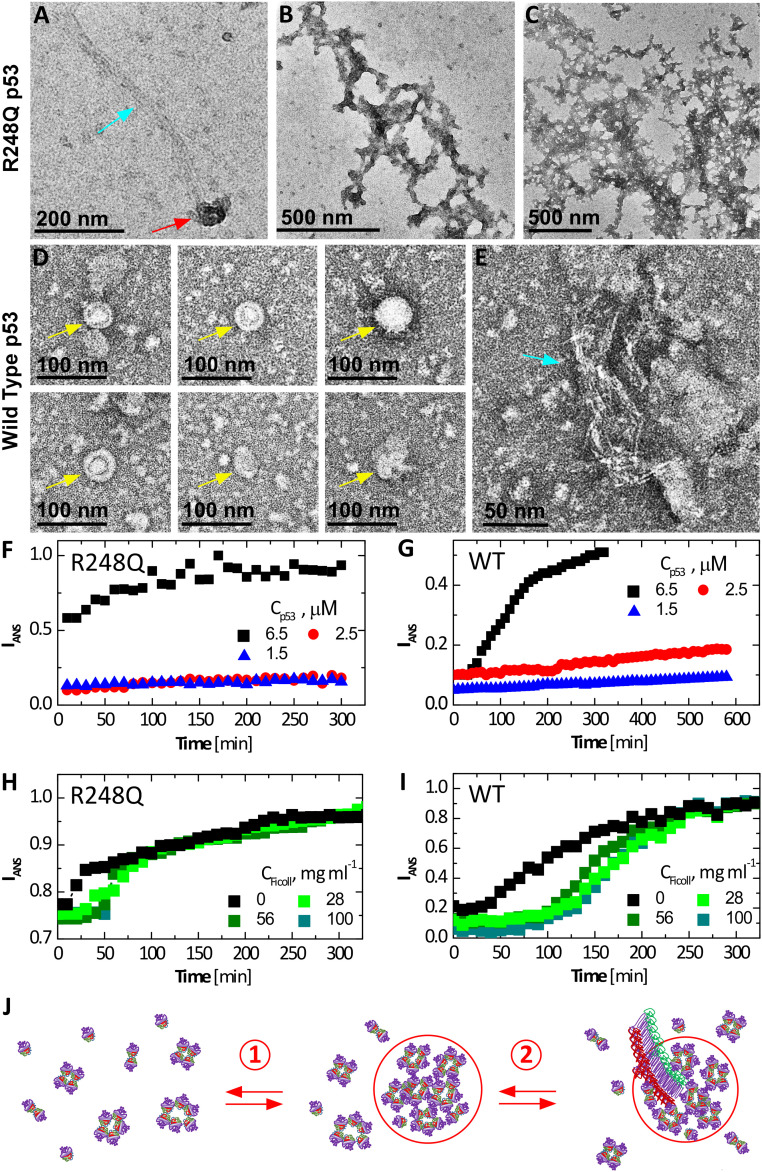
Fig. 3.
Fibrillization of wild type and p53 R248Q. (A–E) Negative staining EM micrographs reveal clusters, fibrils, and amorphous agglomerates of p53 R248Q in A–C and wild-type p53 in D and E. Gold arrows point to empty clusters; red arrows, clusters that spawn fibrils; and blue arrows, fibrils. (B) Branched fibrils coated with amorphous agglomerates. (C) Stand-alone amorphous agglomerates. (F–I) Evolution of the intensity of fluorescence at wavelength 500 nm of ANS in the presence of p53 at 37 °C. ANS concentrations were 200 µM in all tests. (F and G) At the listed concentrations of R248Q, in F, and wild type, in G, in the absence of Ficoll. (H and I) At 6.5 μM of R248Q, in H, and wild type, in I, and in the presence of varying concentrations of Ficoll. (J) Schematic of two-step nucleation of fibrils; step 1) Mesoscopic p53-rich clusters, comprised of misassembled p53 oligomers (depicted here as pentamers), native tetramers, and additional p53 species, assemble and step 2) Fibrils, which likely represent stacks of refolded p53 monomers (7), nucleate within the mesoscopic clusters by the assembly of p53 monomers. Fibril growth proceeds classically, via sequential association of p53 monomers from the solution. Compare to EM micrograph in A.

