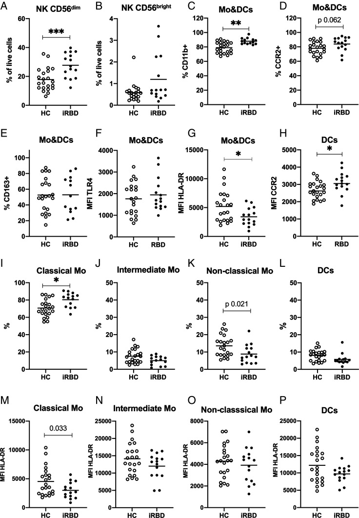
Fig. 1.
Flow cytometry analysis of PBMCs from HCs and iRBD patients. PBMCs in blood from iRBD patients and HCs were analyzed based on TLR2 and CD56 expression. NK cells were identified as TLR2−/CD56+ and divided into (A) CD56 dim mature NK cells and (B) CD56 bright precursor NK cells. TLR2 was used as a pan marker for Mo and DCs and subtyped based on CD14, CD16, and HLA-DR expression. Mo&DCs are shown with respect to the percentage expressing the following surface markers: (C) CD11b (D) CCR2, and (E) CD163; and their MFI of (F) TLR4 and (G) HLA-DR. (H) MFI of CCR2 on DCs. Percentage of the subpopulations in Mo&DCs population: (I) classical monocytes (CD14++/CD16−), (J) intermediate monocytes (CD14++/CD16+), (K) nonclassical monocytes (CD14low/CD16++), and (L) DCs (CD14low/CD16−). The MFI of HLA-DR shown for the Mo&DCs subpopulations: (M) classical Mo, (N) intermediate Mo, (O) nonclassical Mo, and (P) DCs. Lines show means. Mann–Whitney test for B and C, and unpaired t test for A and D–P. *P < 0.05, **P < 0.01, ***P < 0.001. Bonferroni correction (P value set to 0.0125) was applied for I–L and M–P.

