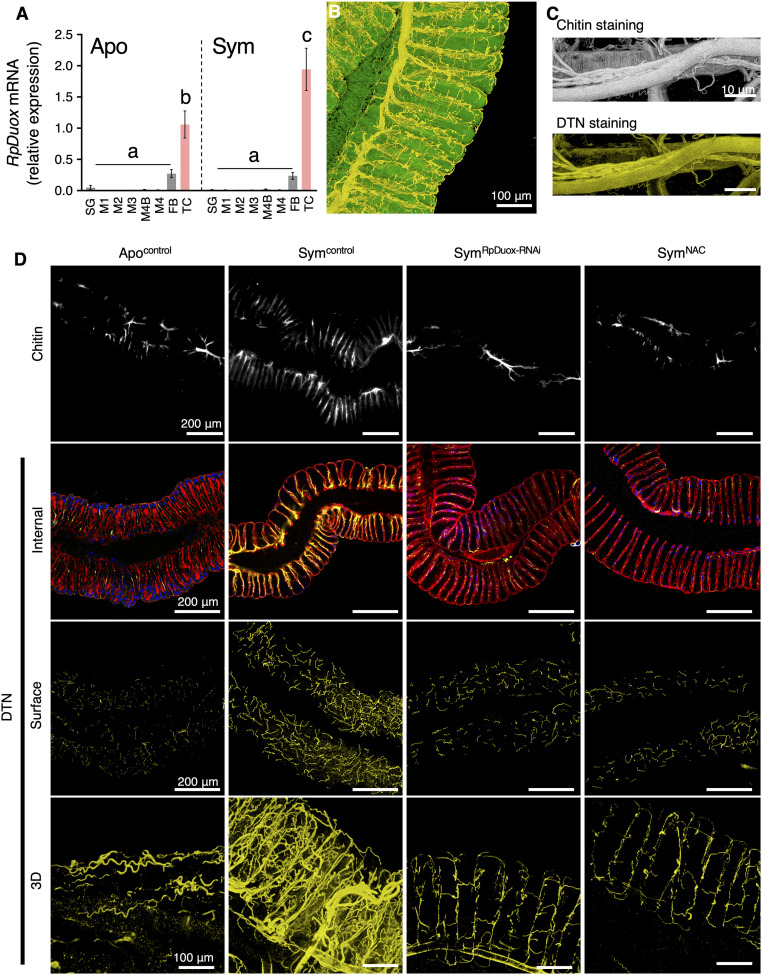
Fig. 3.
Symbiont colonization triggers tracheal development, and RpDuox-mediated ROS stabilizes it by forming a DTN. (A) The relative expression of RpDuox in different tissues of Apo and Sym insects. Data shown are mean ± SD (n = 4 insects). Different letters indicate statistically significant differences (P < 0.05). The statistical significance of differences between samples was analyzed by a Kruskal–Wallis test with Bonferroni correction. (B) A 3D image of the tracheal network in the M4 of a Symcontrol insect. (C) Staining of the chitin and DTN of tracheae detached from the M4. (D) The tracheal network in the M4 of Apo (Apocontrol), Sym (Symcontrol), RpDuox-RNAi (SymRpDuox-RNAi), and NAC-fed (SymNAC) insects. The tracheal network, stained with Fluorescent Brightener 28 (calcofluor-white) to visualize chitin (first row), DTN in the internal section of the M4 (second row), DTN located on the surface of the M4 (third row), and 3D reconstitution of DTN of the tracheal network (fourth row). The colors in B–D are as follows: white, chitin; yellow, DTN; red, host cytoskeleton; blue, host nuclei; green, GFP signal derived from gut-colonizing symbionts.

