Fig. 3.
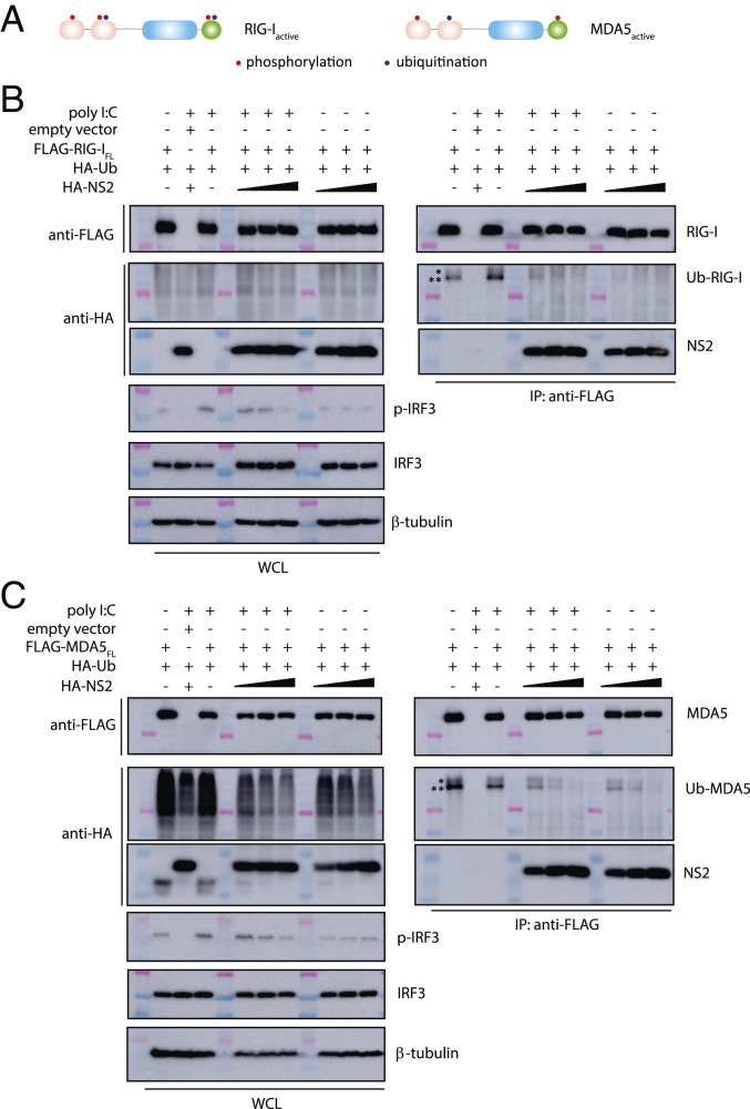
RSV NS2 inhibits RIG-I and MDA5 ubiquitination. (A) Simplified schematic of posttranslational modifications of RIG-I and MDA5. Domains of RIG-I and MDA5 that are phosphorylated (red) or ubiquitinated (blue) are indicated by a single dot. FLAG-coIPs (Right) of FLAG- (B) RIG-IFL or (C) MDA5FL with HA-ubiquitin and increasing concentrations of HA-NS2 (1, 2, and 4 μg) and in the absence or presence of polyI:C transfection. Band corresponding to Ub-RIG-I and Ub-MDA5 is denoted by a double asterisk (**). Unidentified ubiquitinated band is denoted by a single asterisk (*). Immunoblotting of phosphorylated IRF3 (p-IRF3), IRF3, and β-tubulin is shown below. Western blots of WCL are shown on the Left in B and C. Shown are representative results from experiments repeated in triplicate.

