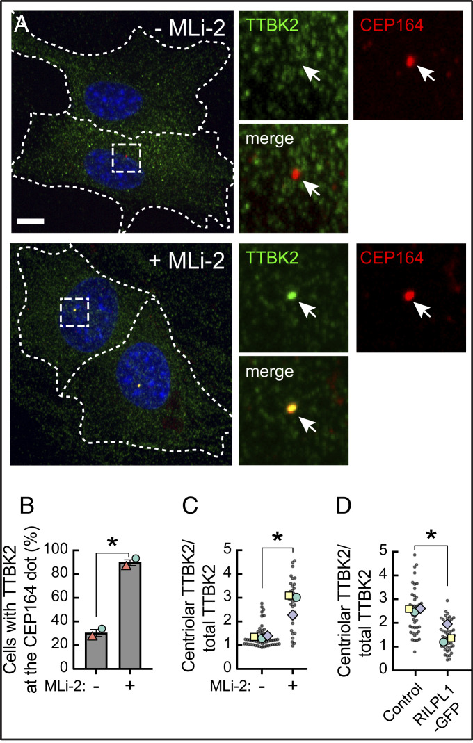
Fig. 6.
Pathogenic LRRK2 activity blocks TTBK2 recruitment without affecting CEP164. (A) R1441C LRRK2 MEF cells stably expressing GFP-TTBK2 were starved for 24 h with 200 nM MLi-2 or DMSO. Cells were then fixed with cold methanol and stained with rabbit anti-TTBK2 (green) and mouse anti-CEP164 (red) antibodies. At right are enlarged regions boxed in the larger images at left. (B) Percent of cells with TTBK2 at the mother centriole, marked by CEP164 staining, by visual inspection. Values represent the mean and SEM of two independent experiments (n > 30 in each experiment). Significance was determined by the paired t test; *P = 0.034. Colored shapes represent the mean of each separate experiment. (C) Intensity of TTBK2 staining over the centriole as a function of its overall expression in cells. Each dot represents the ratio of mean TTBK2 signal intensity at the centriolar area divided by the mean signal intensity throughout the cell (n > 30). Colored shapes represent the mean of each separate experiment. P = 0.036. (D) Effect of RILPL1 expression on TTBK2 levels at the mother centriole. RPE cells were transfected with RILPL1-GFP; its expression was induced with 1 µg/mL doxycycline, and cells were serum starved for 24 h, methanol fixed, and stained for endogenous TTBK2 and CEP164. Cells were then imaged and analyzed as in C. Each dot represents the ratio of mean TTBK2 signal intensity at the centriolar area divided by the mean signal intensity throughout the cell (n > 40). Colored shapes represent the mean of each experiment. Significance was determined by the paired t test, P = 0.034. (Scale bars, 10 µm.)

