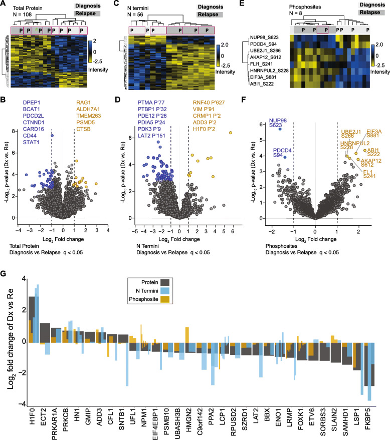
Fig. 3.
PDXs reflect proteome changes that distinguish diagnosis and relapse conditions in patients. a Heatmap on z-score normalized intensities of 108 proteins that significantly distinguished diagnosis from relapse B-ALL conditions (supervised hierarchical clustering, unpaired Student’s t-test, FDR < 0.05). Purple border highlights paired patient (P) and PDX samples. b Fold change and p-value profiles of the top differentiating proteins (FDR < 0.05) in diagnosis and relapse samples. c Supervised hierarchical clustering on 56 N termini that significantly distinguished (unpaired Student’s t-test, FDR < 0.05) diagnosis from relapse samples. Plotted data was z-score normalized. Purple border highlights paired patient (P) and PDX samples. d N termini with significant differences between diagnosis and relapse time-points (FDR < 0.05) are highlighted in a scatter plot. e Supervised hierarchical clustering on 8 phosphosites with differential abundance in diagnosis and relapse cases (ANOVA test, FDR < 0.05, z-score normalized values plotted). Purple border highlights paired patient (P) and PDX samples. f Scatterplot shows significantly dysregulated phoshosites in diagnosis and relapse patients (q value < 0.05). g Fold change (Log2) of 31 proteins differentially regulated proteins (grey bars) in diagnosis compared to relapse samples that also had phosphosites and N termini quantified. Log2 fold change (diagnosis vs. relapse) of associated protein phosphosites (brown bars) and N termini (blue bars) are inserted within the protein bar plots

