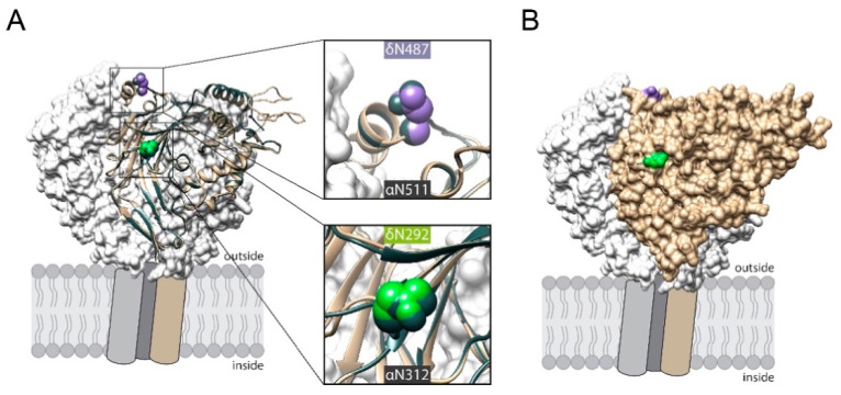
Figure 3.
(A) Cartoon representation of human δ ENaC’s extracellular domain (highlighted in tan) that is aligned to human α ENaC, coloured in slate grey. Extracellular domains of wt β (grey) and γ subunit (white) are shown as surface representation. The predicted localization of the inserted asparagines 292 (green) and 487 (purple) are highlighted together with the corresponding asparagines in α ENaC (N312 and 511, slate grey). (B) Solvent accessible surface representation of δ ENaC indicates that the inserted asparagine residues are localised at exposed positions.

