Figure 1.
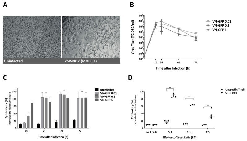
Infection of B16-OVA cells by virotherapy (VSV-NDV) and targeted cytotoxicity by T cells (OTI). (A) B16-OVA cells were infected with VSV-NDV at MOI 0.1 (right panel) or left uninfected (left panel), and images were captured 16-h post-infection. Representative images demonstrating characteristic syncytia formation in the infected well and the healthy uninfected monolayer were captured at 200× magnification. (B) B16-OVA cells were seeded and infected with VSV-NDV (VN) at different MOIs. Virus titers were determined via a TCID50 assay from tissue culture supernatants collected at indicated times after infection. (C) Corresponding cytotoxicity analysis was determined via an LDH detection assay and normalized to a maximum release control. (D) The cytotoxic effector function of OTI T cells on B16-OVA melanoma cells was determined by LDH assay. Cells were co-cultured with OTI or unspecific control T cells for 24 h in the indicated effector-to-target ratios. T cells were isolated from the spleens of OTI or C57Bl6 mice and expanded in vitro before addition to the co-culture. Additional wells of B16-OVA cells were left without the addition of T cells, in order to demonstrate the baseline level of cytotoxicity. All data are presented as mean + SEM of triplicate experiments, and statistical significance was determined by student’s t-test (** p < 0.01, *** p < 0.001).

