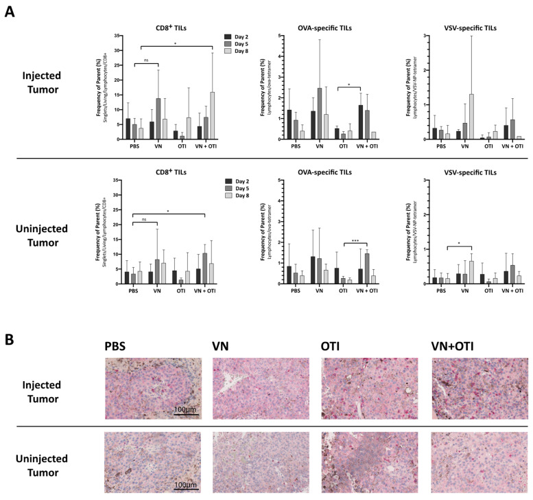
Figure 4.
Features of tumor infiltrating lymphocytes in the injected and uninjected tumor. (A) Mice were subcutaneously implanted with 2.4 × 10⁵ B16-OVA cells on both flanks. One week later, the mice were randomly distributed into indicated treatment groups (n = 3–6) and injected intratumorally with VSV-NDV (VN) at a dose of 107 TCID50 or PBS in an equal volume of 50 µL on day 0, followed by an intravenous OTI T cell injection (5.5 × 10⁶) on day 1 for combination treatment or OTI monotherapy. Tumors from both flanks were harvested on day 2, 5 and 8 after the first treatment. Single cell suspensions were generated from tumor tissues, and tumor infiltrating lymphocytes (TILs) were analyzed for CD8 surface expression and OVA- or VSV-specificity via flow cytometry. Data are presented as mean + SD, and statistical significance was determined by student’s t-test (* p < 0.05, *** p < 0.001). (B) Paraffin-embedded tumor sections of 2 µm in thickness were subjected to immunohistochemical analysis using antibodies specific for mouse CD8 and visualized using a fast red chromogen for detection. Representative sections of injected (top panel) or uninjected (bottom panel) tumors from the indicated treatment groups were captured at 200× magnification. Scale bars indicate 100 µm.

