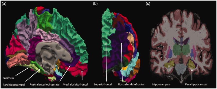
Fig. 1.
Representative MRI images taken using 7-T MRI. (a) Brain structures from the medial aspect view, (b) anterior aspect view, and (c) coronal view of patients with PD in images taken with 7-T MRI and processed with N3 algorithms. The gray matter (GM) thickness of patients with PD was calculated using these algorithms.

