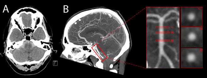
Figure 1.
A-B: (A) Axial noncontrast CT of the head showed diffuse subarachnoid hemorrhage in the posterior fossa suggesting a posterior circulation culprit lesion. (B) Sagittal maximum intensity projection of a CTA head revealed no appreciable aneurysm along the course of the basilar artery. Representative orthogonal planes of the basilar artery lumen are depicted in the insets.

